banner
New Surgical Technique of Calcaneus Fractures - Double Medical Calcaneus Nail System
2021/07/12
Fracture of calcaneum is one of the common fractures of the foot, which is mainly caused by external forces trauma to destroy the bone at the heel and interrupt its intergrity or continuity. The main manifestations of patients are severe pain, swelling, ecchymoses, Heel valgus deformity, flat foot, inability to walk normally.

Calcaneal fractures is one of the most common foot fractures in young adults, accounting for 70% of tarsal bone fractures, 30.3% of all foot fractures, and 2.9% of total body fractures.

The features of Calcaneus Anatomy

1.The calcaneus, the largest tarsal bone of the foot, is an irregular rectangular structure. The structure of that is a thin cortex surrounding the rich cancellous bone.
2.The calcaneus has six facets and four articular surfaces: the anterior, middle and posterior talar articular surfaces. The middle and posterior talar articular surfaces have a wide lateral opening groove, which is called the tarsal sinus;
3.Bohler angle: normal angle is 25°~40°. When the calcaneus is fractured, this angle may decrease, disappear, or become negative, which could affect the posterior wall of the arch of the foot, thereby reducing the strength of the triceps calf.

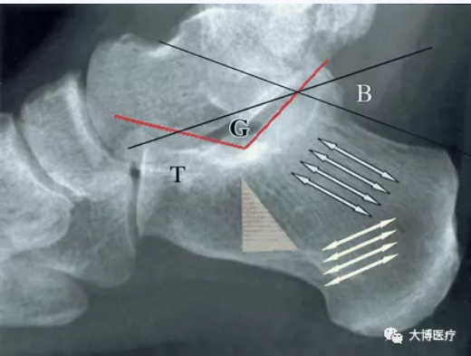
4.Gissane Angle: The foreign statistical data is 120°~145°, and chinese data is 123.8+-8.7°. This Angle is often increased when calcaneus is fractured. And it is used clinically to determine the degree of fracture and evaluated the efficacy.


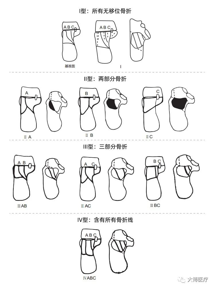
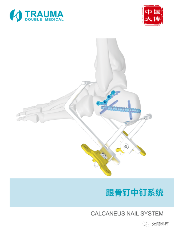
Classification of intra-articular fractures of the calcaneus: Sanders Classification


Clinical Cases:

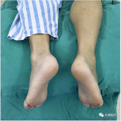
Patients information:

Male, 52, Caused by fall, the right heel is swollen and the movement is limited. Hospital treatment is 2 days.

Auxilliary examination: X-ray: comminuted fracture of right calcaneus.


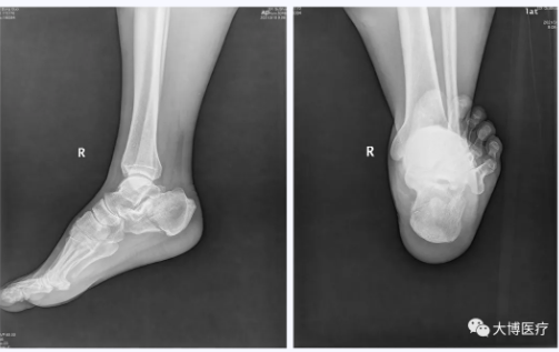
Preoperative Images:


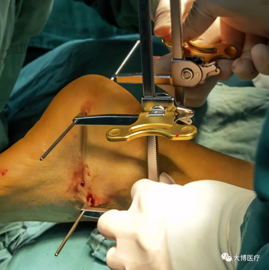
The operation situation:


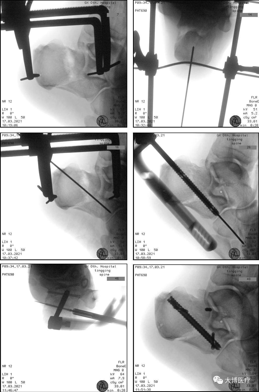
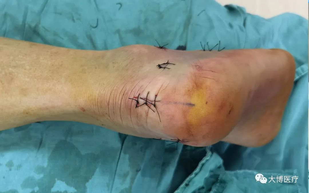
Postoperative images:


Conclusion:

1.The advantages of the traditional open internal fixation with reduction plates: good reduction and stable fixation. But due to the special soft tissue structures of the heel, the incision will be not easy to heal, and the incidence of complications such as surgical site infection is very high.

2.The advantages of the general minimal invasive closed reduction and internal fixation with cannulated nails: Small incision, low infection rate.
But due to the limitations of manual reduction and Kirschner wire pry, it is difficult to achieve a good anatomical reduction. It is also difficult to maintain the stability of bones with cannulated nails alone, which is prone to loss of reduction in the later stage.

3.The advantages of the Calcaneus Cannulated Screw System:

  • Minimal invasive incision: percutaneous minimally invasive incision approach, small soft tissue incision, highly reduce the rate of postoperative complications.
  • Closed reduction: distraction and reduction devices can making distraction and reduction of the length, width and height of the calcaneus; Bilateral distraction prevent calcaneus varus deformity.
  • Accelerated rehabilitation: the interlocking screw in screw structure has stable biomechanical properties, and the subtalar joint locking plate provides strong support and fixation, facilitating early functional rehabilitation training, which is in accord with EARS concepts.


Leave A Message
Leave A Message
If you are interested in our products and want to know more details,please leave a message here,we will reply you as soon as we can.

Home

products

about

contact