Intra-articular calcaneal fractures are usually treated with traditional internal fixation involving a lateral widening incision by the use of
calcaneal locking plates after resetting. While this method allows patients to return to work as soon as possible after the operation, it has several disadvantages. These include a large trauma caused by the L-shaped incision, the risk of skin necrosis, potential damage to the peroneal nerve during the operation, significant postoperative external adhesion, and limited mobility function. To avoid these complications, clinicians have started shifting towards minimally invasive internal fixation techniques.
Minimally invasive internal fixation has proven to be effective in treating patients with intra-articular fractures of the heel bone. It involves smaller incisions, utilizes X-ray fluoroscopy-assisted resetting, facilitates surgical operations, minimizes soft tissue stripping to protect the surrounding blood supply, and provides a favorable biological environment for soft tissue repair and fracture healing. This approach significantly reduces the incidence of adverse reactions and shortens the operation time. In recent years, ankle arthroscopy has also been increasingly used for diagnosing and treating intra- or extra-articular diseases of the foot and ankle. The application of arthroscopic technology in heel bone fractures, assisted by arthroscopy, brings new hope and direction to treatment.
Case Information
I. Patient Profile
Basic information: Mr. Zeng, a 39-year-old male.
Disease description: The patient accidentally fell from a height of 3 meters, resulting in severe pain in his right heel, inability to bear weight, difficulty walking, and a diagnosis of comminuted fracture of the right heel bone through X-ray examination. The patient has no previous history of trauma or surgery and is in good overall health.
Consultation time: July 2023
Treatment Plan: Arthroscopic minimally invasive pin-in-pin internal fixation for right calcaneal fracture via the tarsal sinus approach.
Surgical Procedure
I. First acquaintance with the patient
The patient was admitted to hospital in July 2023 due to right heel pain and limited activity, which had persisted for 4 hours after a fall from a significant height. X-ray examination confirmed a comminuted fracture of the right heel bone.
Preoperative imaging:
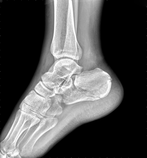
⇑Preoperative DR
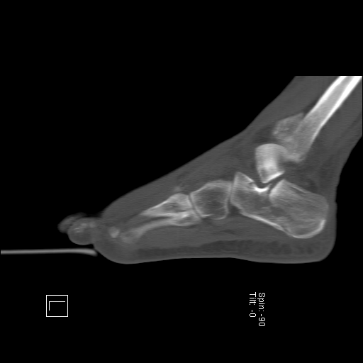
⇑Preoperative CT
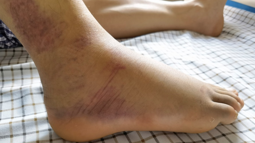
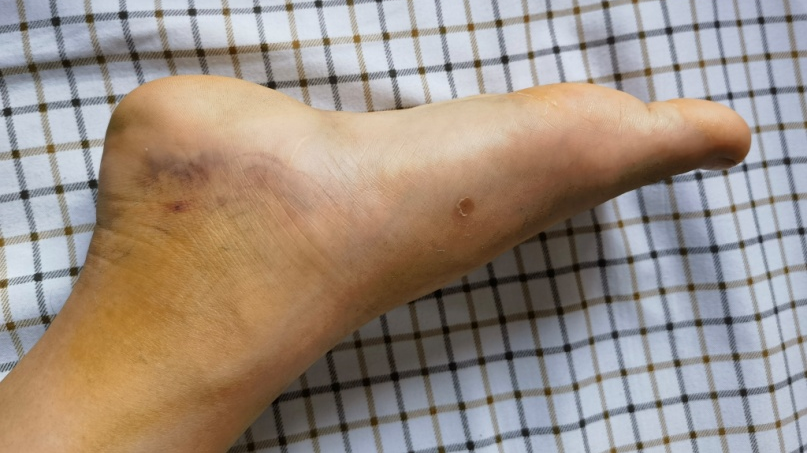
↑ Pre-operative photo
II. Treatment process
Treatment program selection:
The case presented severe collapse of the articular surface following the heel bone fracture, accompanied by significant swelling of the foot and ankle. The following considerations were made:
Conservative treatment was deemed inadequate for resetting the collapsed articular surface. Without proper realignment, the fracture deformity could result in long-term foot and ankle dysfunction, talonavicular joint arthritis, and chronic pain.
Traditional cut-and-replace internal fixation surgery, through the lateral widening approach, offers advantages such as robust internal fixation and reliable resetting. However, it carries the risk of complications, including extensive intraoperative soft tissue stripping, soft tissue damage, postoperative infection, skin edge necrosis, and postoperative adhesions.
Minimally invasive internal fixation was chosen due to its reduced complications and significant efficacy. Arthroscopy allows direct visualization of the collapsed area of the heel bone's posterior articular surface, facilitating monitoring of the resetting effect. The type II heel nail-in-nail system was used to interlock and fixate the posterior articular surface, maintaining the length, height, and width of the heel bone. Minimally invasive surgery requires less swelling of the skin and soft tissues and can be performed at an early stage. Therefore, the surgical plan selected was minimally invasive pin-in-pin internal fixation with arthroscopic restoration of the right heel fracture through the tarsal sinus approach.
Preoperative preparation:
Routine preoperative examinations were conducted, and the affected limb was elevated while ice was applied to reduce swelling. Additionally, measures were taken to prevent thrombosis and other complications.
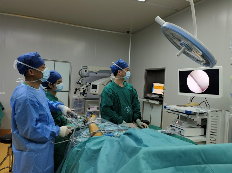
Surgical presentation:
The procedure began with the use of a 2.7mm arthroscope to observe the collapse of the posterior articular surface of the heel bone through the tarsal sinus approach. Following repositioning, the Double Medical heel bone nail-in-nail system was used for fixation.
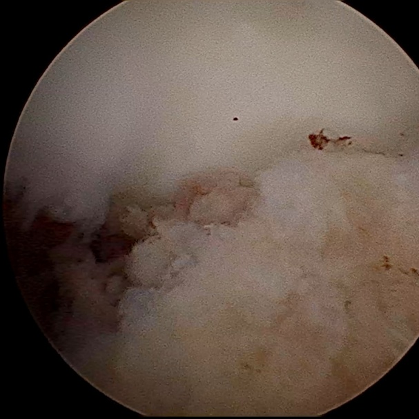
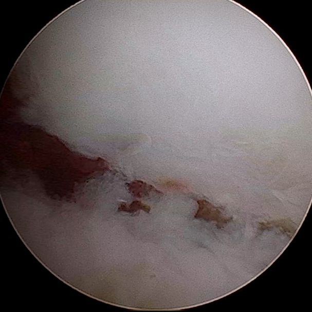
↑ Arthroscopic observation of the collapse of the posterior articular surface of the heel bone
|
Before
|
After
|
|
|
↑ Arthroscopic photos
Postoperative care:
Attention was paid to the wound condition, with the affected limb elevated to reduce swelling, and regular changes of wound dressings.
Gradual functional exercise was emphasized, focusing on active toe movements and primarily engaging in active activities with moderate resistance to prevent muscle atrophy.
Regular radiographic evaluations were performed to monitor fracture healing.
Treatment Effect
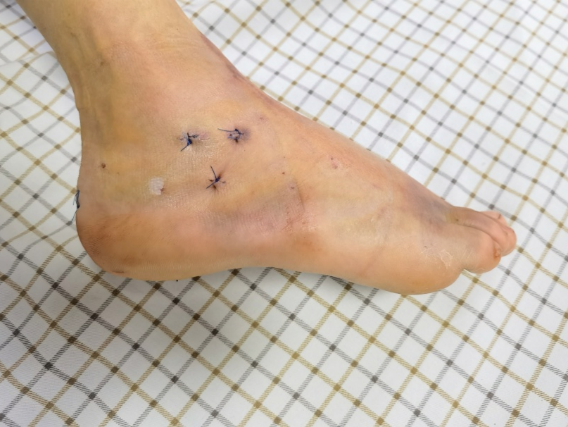
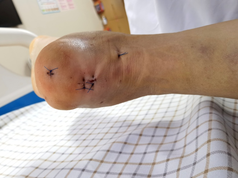
Postoperatively, the patient experienced significant relief from foot pain. The minimally invasive incision was aesthetically pleasing, and the wound remained dry and clean without any signs of oozing. Due to successful fracture resetting, limb swelling subsided noticeably, allowing for early functional exercises of the ankle. The following images display the incision and postoperative progress.
↑ Minimally invasive incision
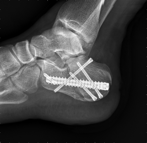
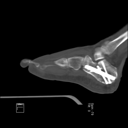
↑ Post-operative DR
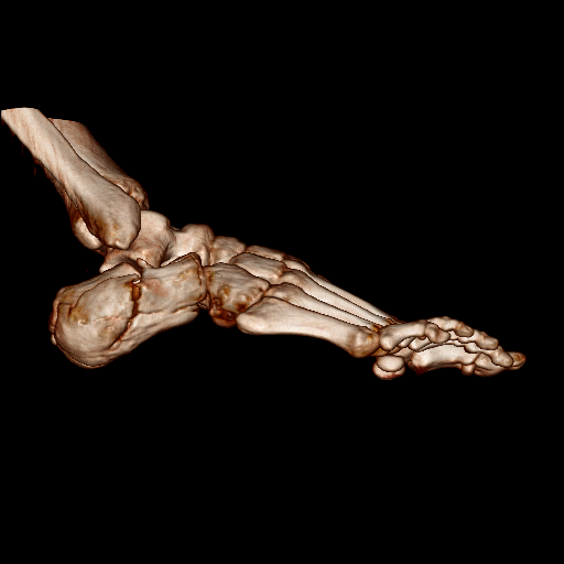
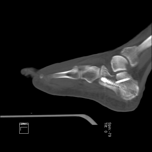
↑ Post-operative CT
IV. Precautions for Patients in Daily Life
Dietary advice: Emphasize a light diet while ensuring intake of nutrients such as protein, vitamins, fiber, calcium ions, and trace elements. Recommended food sources include meat, eggs, milk, vegetables, fruits, fish, shrimp, nuts, among others.
Functional exercise:
Reconstruction of ankle joint mobility: Focus on restoring normal ankle joint movement ability.
Ankle, calf, and plantar muscle strength training: If weight-bearing is permitted, engage in exercises that restore weight-bearing capacity through ankle, calf, and plantar muscle strength training. Specific training methods include plantar pinch marbles, towel grasping exercises, paper clip exercises, and plantar intrinsic muscle training.
Rechecking suggestions: Follow-up appointments should be scheduled at 1, 2, 3 months, six months, and one year after the operation. These visits will help determine fracture healing progress and guide rehabilitation exercises.
Doctor's Perception:
The minimally invasive nail-in-nail internal fixation technique, performed through the tarsal sinus approach and arthroscopic restoration, resulted in minimal damage, fewer complications, restoration of the anatomical shape of the heel bone, and maintenance of fixed heel bone length, height, and width. The interlocking nail-in-nail system provides stable structural and mechanical properties. Overall, the surgery is straightforward, saving operation time, and enabling early functional rehabilitation in line with the Enhanced Recovery After Surgery (ERAS) concept.


 Tel : 86 592 6087101
 Email : info@double-medical.com