

Distal tibial fractures can be challenging to treat because the distal tibia has limited soft-tissue coverage and is located close to the ankle joint. Treatment decisions depend on the fracture pattern, degree of displacement, articular involvement, bone quality, and condition of the surrounding soft tissues.
For selected extra-articular and metaphyseal fractures, intramedullary nailing can provide stable fixation while limiting additional disruption of the soft-tissue envelope. Modern distal tibial nail systems are designed with distal locking configurations that help stabilize the relatively short distal fragment.
This article claim that the main treatment options for distal tibial fractures, when intramedullary nailing may be considered, and the fixation principles surgeons should evaluate when selecting a distal tibial nail.
What You Need to Know
- Intramedullary nailing is an established surgical option for selected distal tibial fractures, particularly extra-articular metaphyseal fractures when adequate distal bone is available for stable interlocking fixation.
- Treatment selection depends on fracture location, displacement, articular involvement, soft-tissue condition, bone quality, and the amount of distal bone available for fixation.
- Plate fixation, external fixation, and intramedullary nailing each have a role in distal tibial fracture treatment, depending on the individual injury.
- Modern distal tibial nail systems use multiple distal locking screws to improve stability in the short distal tibial segment.
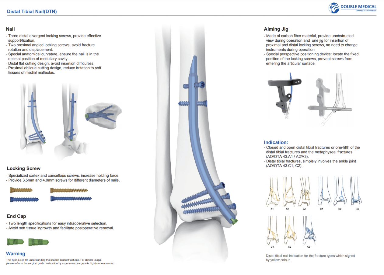
- The Distal Tibial Nail from Double Medical is designed for selected closed and open distal tibial fractures and metaphyseal fractures, with three distal divergent locking screws, two proximal angled locking screws, and a special anatomical curvature.
What Is a Distal Tibial Fracture?
A distal tibial fracture is a break in the lower portion of the tibia near the ankle. These injuries range from relatively simple extra-articular fractures to complex fractures extending into the ankle joint, including pilon fractures.
The location and morphology of the fracture are important when selecting a fixation method. Fractures close to the tibial plafond may provide only a short distal segment for fixation, making stable implant positioning more challenging. AO guidance notes that fractures located very close to the articular surface may not have sufficient distal bone for reliable intramedullary nail fixation.
Why Are Distal Tibial Fractures Difficult to Treat?
The distal tibia has relatively limited soft-tissue coverage compared with other regions of the lower leg. As a result, swelling, fracture blisters, open wounds, and other soft-tissue injuries can influence both the timing and choice of surgical fixation.
The injury mechanism can also vary. High-energy trauma, such as motor vehicle accidents or falls from height, can produce displaced or comminuted fractures with significant soft-tissue damage. Lower-energy twisting injuries may produce less severe fracture patterns, while falls in older patients with reduced bone density can also result in distal tibial fractures.
For these reasons, radiographic findings should be considered together with the patient’s overall condition and soft-tissue status when planning distal tibia fracture surgical treatment.
Distal Tibial Nail (DTN)
Overview of Distal Tibial Fracture Treatment Options
The appropriate treatment for a distal tibial fracture depends on the fracture pattern and patient-specific factors. Management may be non-surgical or surgical.
Non-Surgical Management: Casts and Braces
Stable fractures with acceptable alignment and minimal displacement may sometimes be treated without surgery. Immobilization with a cast or brace can help maintain alignment while the fracture heals.
However, displaced or unstable fractures may require surgical fixation to restore and maintain alignment.
Surgical Treatment for Displaced Distal Tibial Fractures
Several fixation strategies may be considered.
Plate Fixation
Plate and screw fixation can provide direct control of fracture alignment and is particularly useful when precise reconstruction of the articular surface is required.
For complex intra-articular injuries such as pilon fractures, fixation may involve direct or indirect articular reconstruction combined with appropriate metaphyseal fixation.
Minimally invasive plate osteosynthesis (MIPO) may also be considered in selected extra-articular fractures when preservation of the soft-tissue envelope is an important consideration.
External Fixation
External fixation may be used for temporary or definitive stabilization depending on the injury. It can be particularly useful when there is substantial swelling, severe soft-tissue injury, or an open fracture requiring staged management.
Intramedullary Nailing
Intramedullary nailing provides internal fixation through the medullary canal rather than requiring extensive exposure of the fracture site.
For appropriately selected distal tibial fractures, a nail can provide load-sharing fixation while limiting additional disruption to the surrounding soft tissues. However, the nail must have sufficient distal fixation capacity because the distal tibial fragment can be relatively short. AO guidance recommends adequate distribution of distal interlocking screws to provide sufficient stability.
When Is Intramedullary Nailing Indicated for Distal Tibial Fractures?
The key question is not simply whether an intramedullary nail can be used, but when an intramedullary nail is an appropriate fixation option for the specific fracture pattern.
Primary Indications for Distal Tibial Intramedullary Nailing
Intramedullary nailing may be considered for:
- Extra-articular distal tibial metaphyseal fractures
These fractures, including selected AO/OTA 43-A fracture patterns, are among the main indications for distal tibial intramedullary fixation. The fracture should leave sufficient distal bone for stable interlocking fixation.
- Displaced or unstable fractures where soft-tissue preservation is important
Because intramedullary nailing can be performed without extensive exposure of the fracture site, it may be considered when the surrounding soft-tissue envelope makes extensive plating less desirable.
This can be particularly relevant in high-risk patients or fractures associated with significant soft-tissue injury. A 2024 retrospective study reported the use of retrograde tibial nailing in selected high-risk patients with extra-articular distal tibial fractures, including patients with conditions such as severe osteoporosis and soft-tissue compromise.
- Selected fractures with limited, non-displaced articular extension
Some fractures with an articular component may still be treated with intramedullary nailing when the articular extension is suitable for additional fixation.
AO guidance notes that selected fractures with undisplaced articular extensions may be treated with intramedullary nailing after the articular fracture line has been stabilized appropriately, such as with lag screws.
- Selected open distal tibial fractures
Intramedullary fixation may be considered in selected open fractures as part of an overall treatment strategy. The decision depends on contamination, soft-tissue condition, fracture configuration, and the need for debridement or staged management.
- Selected patients where an alternative entry point may be advantageous
Traditional antegrade tibial nailing uses a proximal tibial entry point and can be associated with anterior knee symptoms. Retrograde techniques use a distal entry point and may therefore be considered in selected distal tibial fractures where avoiding a proximal tibial entry point is advantageous.
Recent comparative research has specifically evaluated retrograde versus antegrade intramedullary nailing for extra-articular distal tibial fractures, suggesting that retrograde nailing may be a useful option in appropriately selected patients.
DTN Distal Tibial Nail for Selected Distal Tibial Fractures
For suitable fracture patterns, implant design becomes particularly important because the distal tibial fragment may provide limited space for fixation.
The DTN Distal Tibial Nail from Double Medical is indicated for selected closed and open distal tibial fractures and metaphyseal fractures, including AO/OTA 43.A1, 43.A2, and 43.A3 patterns. The system also lists selected distal tibial fractures with simple ankle-joint involvement, including AO/OTA 43.C1 and C2 patterns.
Its fixation design includes:
- Three distal divergent locking screws for distal support and fixation
- Two proximal angled locking screws to help resist fracture rotation and displacement
- Special anatomical curvature designed to position the nail appropriately within the medullary cavity
- Distal flat cutting design to facilitate nail insertion
- Proximal oblique cutting design intended to reduce soft-tissue irritation around the medial malleolus
These features address an important principle of distal tibia fracture fixation: stable fixation requires sufficient distal screw number, appropriate screw distribution, and accurate fracture reduction. AO guidance similarly emphasizes the importance of having multiple well-distributed distal interlocking screws in the short distal tibial segment.
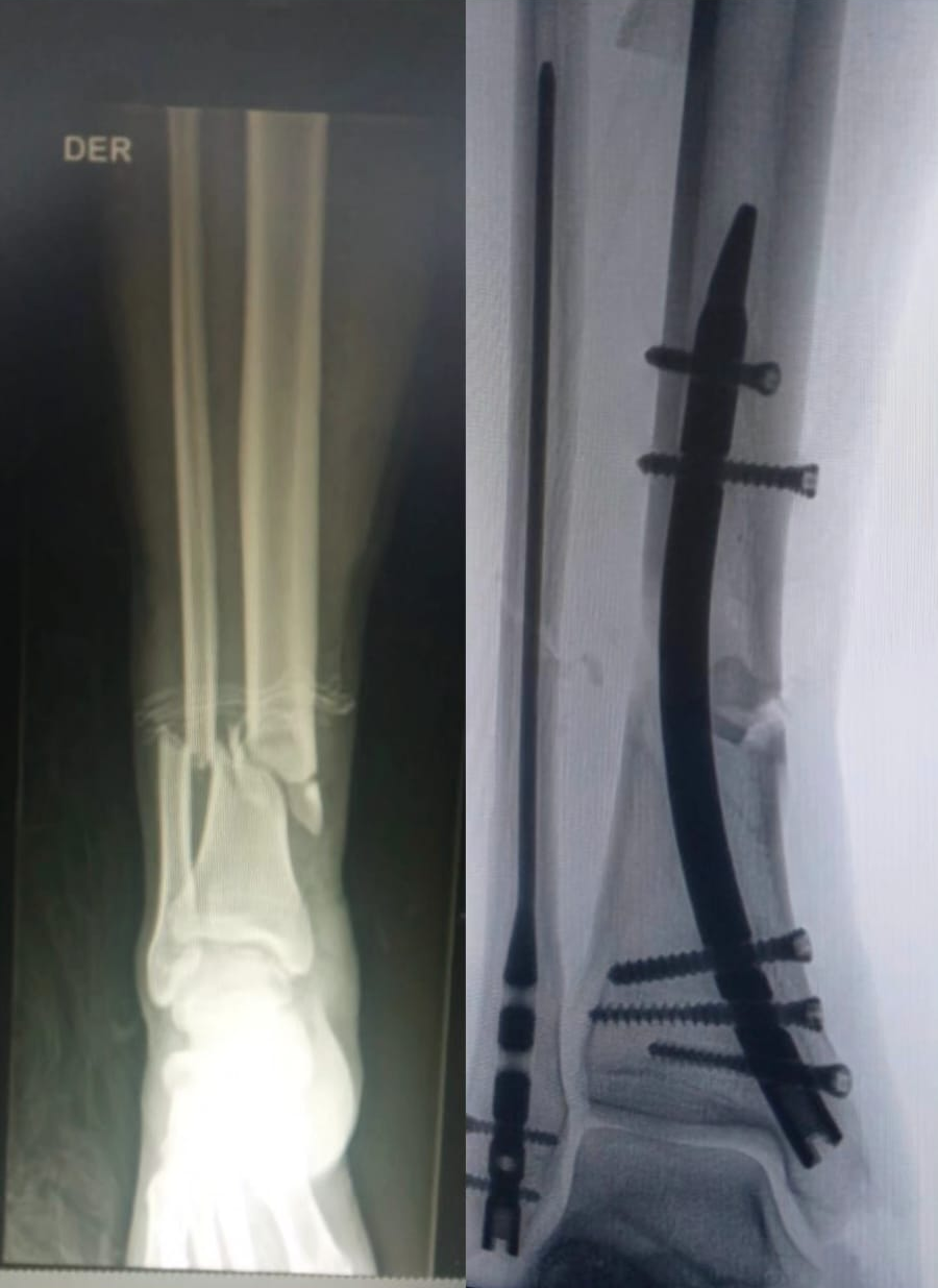
Case_DoubleMedical’s DTN
When Is Intramedullary Nailing Not Recommended?
Intramedullary nailing is not appropriate for every distal tibial fracture. Careful assessment of the fracture and the available distal bone stock is essential.
Potential situations where another fixation strategy may be more appropriate include:
Complex Intra-Articular Pilon Fractures
Severely comminuted pilon fractures may require direct reconstruction of the ankle joint surface. In such cases, a fixation strategy that provides direct control of the articular fragments may be preferred.
Fractures Too Close to the Articular Surface
If the fracture is located extremely close to the tibial plafond, there may not be enough distal bone to accommodate an adequate number and distribution of interlocking screws.
AO guidance specifically notes that fractures within approximately 1–2 cm of the articular surface may not be adequately stabilized with an intramedullary nail.
Severe Bone Loss or Contamination
Extensive bone loss, severe contamination, or soft-tissue conditions requiring staged treatment may make immediate intramedullary fixation inappropriate.
Skeletally Immature Patients
Standard adult distal tibial nail systems are generally not intended for patients with open growth plates. Pediatric fracture management requires consideration of the physis and age-specific fixation strategies.
How Does It Work?
The term retrograde distal tibial nailing generally refers to introducing the nail from the distal tibia and advancing it proximally through the medullary canal.
This differs from traditional antegrade tibial nailing, which uses a proximal entry point near the knee.
Retrograde vs. Antegrade Tibial Nailing
The choice between retrograde and antegrade nailing depends on fracture location, implant design, surgical technique, and patient-specific factors.
A potential advantage of a distal entry approach is that it avoids the proximal tibial entry point used for conventional antegrade tibial nailing. This may be relevant for patients in whom anterior knee symptoms are a concern.
However, retrograde nailing does not eliminate all potential postoperative pain or complications. Distal entry-site irritation, screw prominence, malalignment, and other complications remain possible and should be evaluated during surgical planning.
A 2026 study of retrograde tibial nailing for extra-articular distal tibial fractures reported fracture healing in its patient cohort while also investigating postoperative anterior ankle pain, illustrating the importance of evaluating both fixation stability and entry-site considerations.
Key Surgical Principles
The exact surgical technique varies according to the implant system and fracture pattern, but important principles include:
- Assess the soft tissues before definitive fixation
Significant swelling, fracture blisters, and open wounds may influence the timing and type of fixation.
- Achieve and maintain fracture reduction
Correct alignment should be established before final nail insertion and confirmed using appropriate intraoperative imaging. AO guidance emphasizes maintaining reduction during nail insertion and confirming alignment fluoroscopically.
- Ensure adequate distal fixation
Because the distal tibial segment can be short, distal locking screws should provide sufficient support and appropriate distribution around the nail.
- Control rotation and translation
Distal tibial fractures can be susceptible to malalignment and rotational deformity. The locking configuration, reduction technique, and implant design all contribute to construct stability.
- Consider associated fibular and ankle injuries
Associated fractures should be assessed individually. Whether additional fibular fixation is beneficial depends on the fracture pattern and overall stability; recent research continues to evaluate this question.
Clinical Benefits and Potential Risks
Potential Benefits of Intramedullary Nailing
For appropriately selected distal tibial fractures, intramedullary nailing may offer several potential advantages:
Reduced disruption of the soft-tissue envelope
Unlike traditional open plating, intramedullary fixation can limit direct exposure of the fracture site.
Load-sharing fixation
The nail is positioned within the medullary canal, allowing the implant and bone to share mechanical loads.
Multiple options for distal locking
Modern distal tibial nails can provide multiple locking points to improve fixation of short distal fragments. This is particularly important in metaphyseal fractures.
Potential for earlier functional rehabilitation
Stable fixation can facilitate postoperative mobilization and rehabilitation when permitted by the fracture pattern, bone quality, soft-tissue condition, and surgeon’s protocol.
Potential Risks and Complications
As with other surgical fixation methods, intramedullary nailing carries potential risks, including:
- Malalignment or malreduction
- Rotational deformity
- Screw malposition or irritation.
- Soft-tissue irritation at the entry site
- Infection
- Delayed union or nonunion
- Implant failure.
- Ankle or surrounding joint stiffness.
These risks depend on the fracture pattern, patient factors, surgical technique, implant selection, and postoperative management. Intramedullary nailing should therefore be considered as one treatment option rather than a universally preferred solution.
Postoperative Recovery After Distal Tibial Intramedullary Nailing
Recovery following distal tibial fracture surgery varies considerably between patients.
Factors influencing healing include:
- Fracture severity and location
- Open versus closed injury
- Soft-tissue damage
- Bone quality
- Associated fibular or ankle injuries
- Reduction and fixation stability
- Patient age and general health
Radiographic monitoring is typically used to evaluate fracture alignment and progression toward union.
Weight-bearing is also individualized. Some patients may begin protected or partial weight-bearing according to the stability of the fixation and the treating surgeon’s protocol, while more complex fractures may require a longer period of protection.
Physical therapy can help restore ankle range of motion, lower-limb strength, gait, and functional mobility as healing progresses.
Clinical reports of retrograde intramedullary nailing for selected extra-articular distal tibial fractures have reported fracture union within several months, but individual recovery times vary and should not be interpreted as a universal healing timeline.
Conclusion:
Intramedullary nailing is an important option in the surgical treatment of selected distal tibial fractures, particularly extra-articular metaphyseal fractures with sufficient distal bone for stable interlocking fixation.
The decision to use a distal tibial nail should consider more than the X-ray alone. Fracture location, articular involvement, soft-tissue condition, bone quality, distal bone stock, and the required locking configuration all influence the choice of fixation.
For suitable fracture patterns, modern distal tibial nail systems can provide minimally invasive intramedullary fixation with multiple distal locking options. The Double Medical DTN Distal Tibial Nail is designed for selected closed and open distal tibial fractures and metaphyseal fractures, with features including three distal divergent locking screws, two proximal angled locking screws, and anatomical curvature.